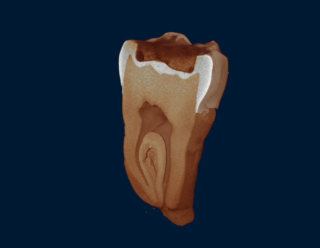
A root canal is not the single straight tube that textbook diagrams like to draw. It is a three-dimensional system that branches, curves, bridges across itself, and sometimes merges into a C-shaped trough. The difficulty is that all of it hides inside dentine, invisible from the outside — and the traditional way to look was to destroy the tooth.
Why this has to be studied population by population
Canal anatomy is not the same the world over. The clearest example is the C-shaped canal in the mandibular second molar, reported anywhere from about 2.7 percent to 48.7 percent depending on which population was studied. Even within South and Southeast Asia the spread is wide — the Burmese population sits at 22.4 percent, markedly above Indian, Thai, and Sri Lankan figures.
A range that wide means findings from one population cannot stand in for another. A clinician treating Thai patients needs to know what Thai teeth actually do, because anatomy nobody expected is a leading reason root canal treatment fails.
What Thai research has already established
- The classic study by Gulabivala and colleagues in 2002 in the International Endodontic Journal examined Thai mandibular molars and found C-shaped roots in 10 percent of 60 mandibular second molars.
- A Chulalongkorn University CBCT study of 1,159 Thai patients found most mandibular premolars are Vertucci type I — 63.1 percent in the first and 98 percent in the second — with over 98 percent single-rooted. Bifurcation appeared in 28.5 percent of first premolars and trifurcation in 3.2 percent.
- Another Chulalongkorn study put MB2 canal prevalence in maxillary molars at roughly 60 percent in first molars and 30 percent in second molars, commonly appearing bilaterally.
- A Mahidol University study examined tooth length and canal morphology across 348 premolars from an indigenous Thai population.
That MB2 figure of around 60 percent has a direct clinical meaning: in more than half of Thai maxillary first molars there is a fourth canal hiding. Miss it when the chamber is opened and it stays uncleaned — a reservoir of infection that surfaces later as a failed treatment.
CBCT and micro-CT answer different questions
Notice that nearly all the Thai work above used CBCT — which suits that question exactly, because it can be collected from live patients and reach sample sizes in the thousands. But CBCT has a ceiling, and that is where micro-CT comes in.
| Aspect | CBCT | Micro-CT |
|---|---|---|
| Used on | Live patients, in the mouth | Extracted teeth in the lab |
| Resolution | Coarser — enough to count canals | Micron level — canal walls and cracks visible |
| Sample size | Hundreds to thousands | Tens of teeth, but far deeper data per tooth |
| Best for | Epidemiology and treatment planning | Testing instruments, techniques, and materials |
The method-changing part: each tooth is its own control
Before micro-CT, seeing what a shaping file does to dentine meant sectioning the tooth or clearing it chemically and looking through it. Both destroy the specimen, so before and after could never be the same tooth — you compared one tooth against another.
The trouble is that teeth differ enormously to begin with — curvature, dentine thickness, canal size. The variation between specimens is often larger than the effect being tested, so researchers had to inflate sample sizes just to see through the noise.
Micro-CT removes the problem entirely. Scan the tooth, prepare the canal by whatever technique is under test, scan it again, then co-register the two datasets into a colour-coded 3D model showing exactly where dentine was removed. Each tooth becomes its own control, between-specimen variation drops out of the equation, and a smaller sample now supports a firmer conclusion than a larger one did before.
What can be measured
- Canal volume, surface area, and perimeter before and after preparation, plus diameter change slice by slice down the canal.
- The proportion of canal wall the instrument never touched — a direct measure of how complete the cleaning was, and something the old methods could barely quantify.
- Canal transportation away from the original axis and centring ability, which matter most in curved canals.
- Dentinal microcracks arising after preparation.
- Retreatment work — what percentage of the old filling material came out, and where the remnants sit.
This kind of work already happens in Thailand. Chulalongkorn University has used micro-CT to compare three supplementary cleaning protocols in teeth root-filled with a bioceramic sealer and then retreated — a question CBCT simply cannot answer.
Where NEOSCAN fits
A tooth is a very small specimen next to what these systems can hold, so size is barely a constraint. The remaining question is how fine you need to see.
| Model | Resolution | Which dental work it suits |
|---|---|---|
| N70 | Under 4 µm / 2.5 µm voxel | General canal morphology and before-and-after comparison — most published work in this field runs at ten to twenty microns, so this is already ample. |
| N80 | 2 µm / submicron voxel | Work that needs dentinal microcracks or the interface between filling material and canal wall, with phase contrast available for tissues of similar density. |
| N90 | 300 nm / 40 nm voxel | Work reaching dentinal tubules or enamel microstructure, with the built-in micro-XRF module for elemental mapping in mineralisation studies. |
The advantage that matters most: it sits on a bench
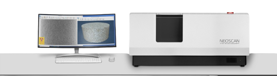
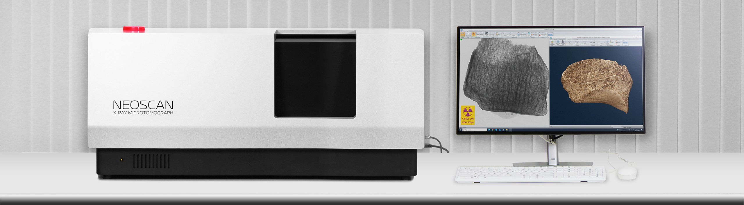
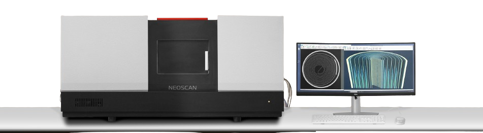
High-resolution industrial X-ray CT usually means a room-sized installation: a purpose-built shielded enclosure, its own power and cooling, and often a place in a central facility where time has to be booked. NEOSCAN goes the other way, compressing lab-grade capability into a system that sits on an ordinary bench.
That sounds like a question of floor space, but its real effect is on method. Before-and-after work means scanning the same tooth at least twice, and three or more times when several treatment steps are compared. If every pass means packing the specimen, shipping it, queueing, and waiting for results, the question stops being askable — so researchers quietly redesign around single-scan studies instead.
- It lives in your own department, so repeat scans are simply available and postgraduate students can actually reach the instrument instead of waiting for a shared facility.
- Specimens never travel — which matters when a tooth has to be repositioned identically each pass for the before-and-after datasets to co-register accurately.
- Budget and footprint sit at a scale a single department can carry, rather than a university-level project that waits years for approval.
- Acquisition, reconstruction, and 3D visualisation come in one platform with free updates, so there is no separate analysis licence to buy.
Put another way, having the scanner in the department does not just make the work more convenient — it changes which questions get asked at all. That is precisely what Alexander Sasov, who founded NEOSCAN, set out to do when he pioneered benchtop micro-CT in its earliest days.
Limits to plan around
- Extracted teeth only. The radiation dose is far too high for a patient, so if the question needs live subjects, CBCT remains the only answer.
- High-resolution scans take hours per tooth, and before-and-after work needs at least two of them. Put machine time into the project plan from the start.
- The datasets are large. Plan the workstation and storage in advance rather than discovering the shortfall later.
- Someone has to know the analysis software and segmentation. The scanner hands you a volume, not a finished number.
- Using extracted human teeth in research requires ethics-committee approval under your institution’s rules.
If you are planning work in this area, the figures worth bringing are: tooth types and how many per project, the smallest detail you must resolve, how many scan passes per specimen, and the analysis software your team already uses. The PMC team can size the system from there.