NEOSCAN
Belgian company founded by microCT pioneer Alexander Sasov, providing tools for non-destructive 3D microscopic measurement.

Specifications & options
Technology
Spatial resolution
300 nm to 2 µm
X-ray source
Up to 160 kV
Applications
Why choose NEOSCAN?
Submicron resolution
Resolve fine internal structures with spatial resolution down to 300 nm and voxel sizes reaching 40 nm.
Non-destructive imaging
Capture full 3D internal geometry without cutting or damaging the sample.
Benchtop footprint
Deliver lab-grade CT performance from a compact system that fits on a standard benchtop.
Integrated software
Acquire, reconstruct, and visualize 3D data in one all-in-one platform with free updates.
Product series
Understand each series to find the right fit for your application.
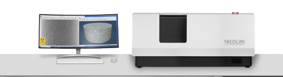
NeoScan N80
Scientific-grade micro-CT scanner with a 2 µm focal spot, sealed 110 kV source, and submicron pixel size for high-resolution 3D imaging.
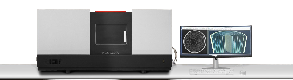
NeoScan N90
A benchtop nano-CT scanner combining a 160 kV transmission source down to 40 nm voxels, dual-detector switching, and an integrated micro-XRF module for elemental mapping.
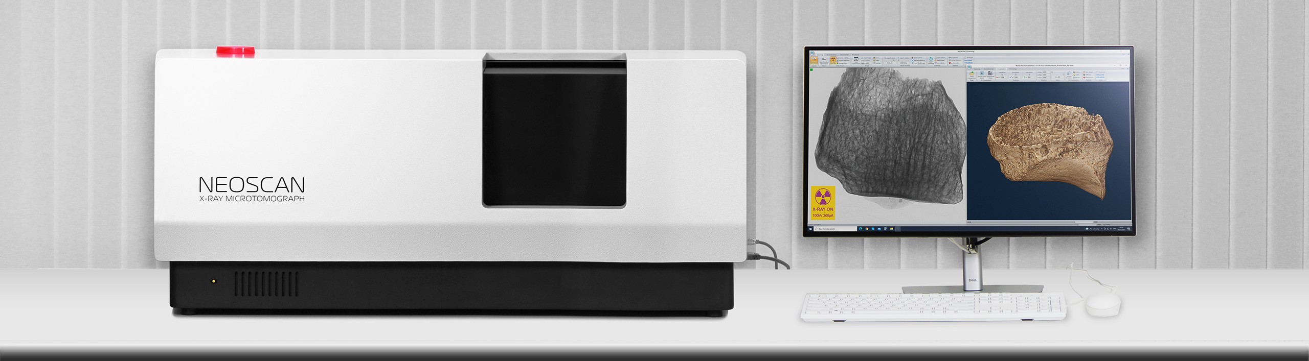
NeoScan N70
A compact benchtop micro-CT scanner for non-destructive 3D internal imaging — suited to research and routine inspection.
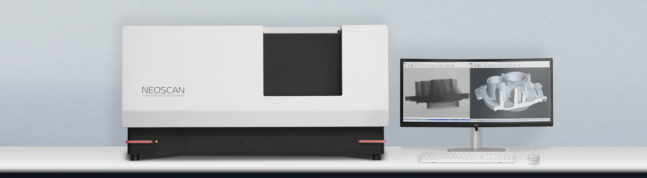
NeoScan NXL
A micro-CT scanner built for larger samples and a wider work envelope, for non-destructive 3D measurement in materials and industrial applications.
Applications
Real micro/nano-CT scan examples across industries.
Advanced GPU↗
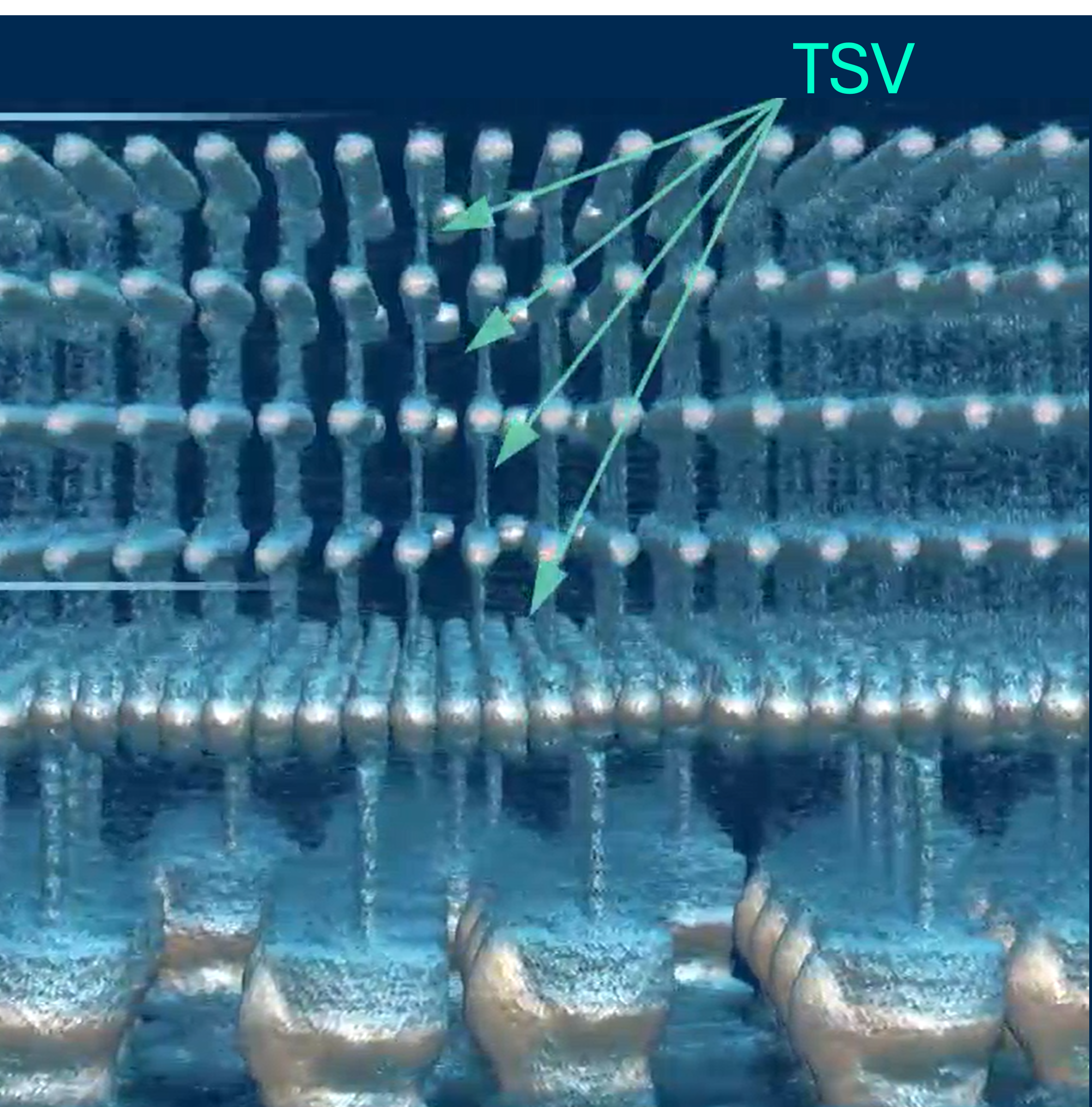
Inspect chip packaging, solder joints, and internal interconnects of high-density processors without desoldering.
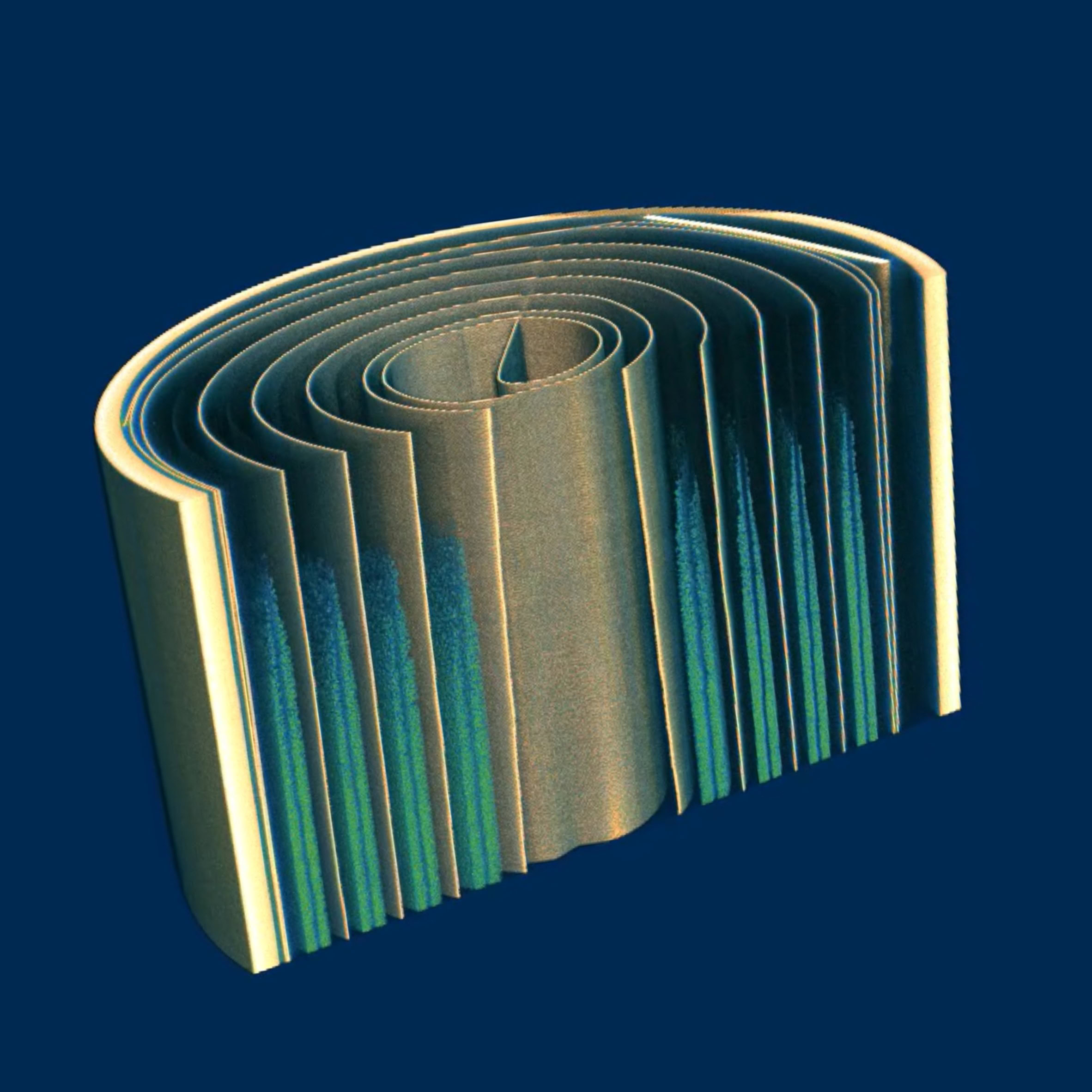
Li-ion Battery↗
Reveal electrode winding, separator alignment, and internal defects in cells for battery R&D and failure analysis.
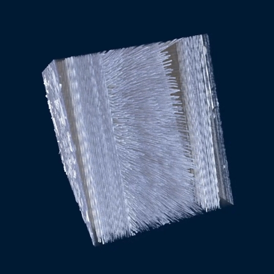
Carbon-fiber Composite↗
Measure fiber orientation, voids, and delamination in CFRP parts to verify structural quality.
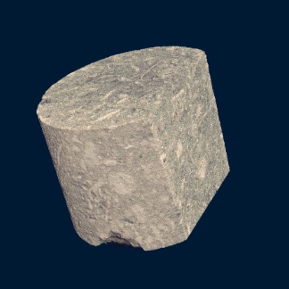
Carbonate Reservoir↗
Visualize pore networks and grain structure in reservoir rock for petrophysics and core analysis.
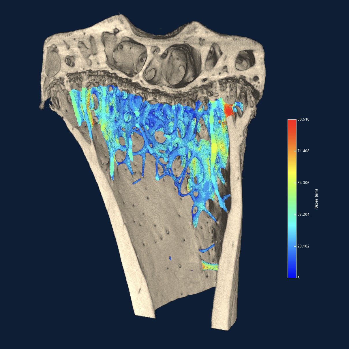
Mouse Bone↗
Quantify trabecular bone structure and density in preclinical research, fully non-destructively.

Molar Tooth↗
Examine enamel, dentin, and internal canals in 3D for dental research and caries studies.
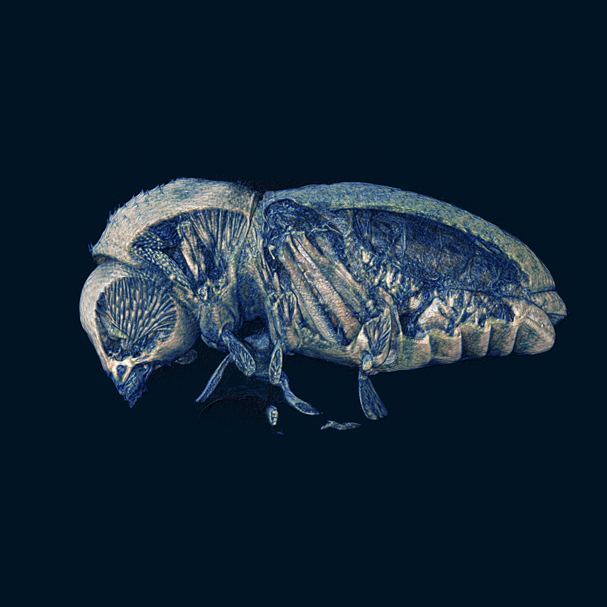
Beetle↗
Capture the full internal anatomy of small organisms for taxonomy and natural-history research.
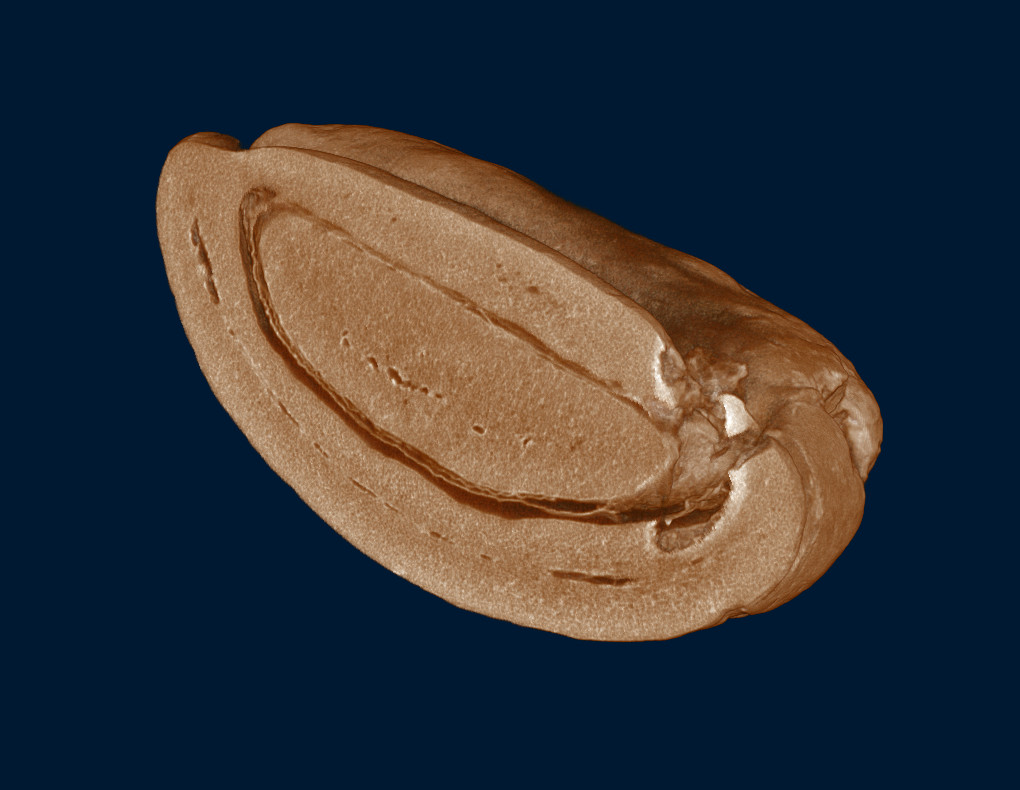
Coffee Bean↗
Study internal porosity and cell structure of beans and seeds for food and agricultural research.
Need a specific model or spare part?
Chat with our team on LINE and we’ll help you find the right one.